Thursday, August 31, 2006


overflow incontinence

Overflow Incontinence ( kan het gevolg zijn van Calcium blockers.)

Overflow incontinence accounts for 10-15% of urinary incontinence. Overflow leakage of urine occurs when there is a mechanical or functional obstruction of the urinary bladder outlet. The obstruction leads to overfill of the bladder and incontinence due to a detrusor contraction which occurs when a certain volume is reached. In this form of chronic incontinence, the client usually does not know why she/he leaks urine and frequent dribbling is common. Often the sensation of bladder fullness is diminished and the stream of urine is weak. These symptoms occur secondary to an anatomic obstruction (i.e. enlarged prostate, urethral stricture) or an atonic bladder. An atonic bladder can result from neurologic injury, diabetic neuropathic bladder, or drug-induced atonia. Drug induced atonia can be caused by anticholinergics, narcotics, anti-depressants, and smooth muscle relaxants. Neurologic injury can be caused by spinal cord trauma or a stroke.


overactive bladder

Evaluation and Diagnosis

OAB is a syndrome which is diagnosed on the basis of history, examination, and urinalysis. The history should focus on the patient's bladder complaints. Does the patient have the symptoms described in the definition of OAB; namely urinary urgency, frequency, nocturia, and UUI? For patients with incontinence it is important to distinguish leakage associated with UUI compared with leakage associated with SUI (Table 1).

Table 1. Differentiating OAB From Stress Incontinence

SymptomsOABStress Incontinence
Urgency (strong, sudden desire to void)YesNo
Frequency with urgencyYesRarely
Leaking during physical activity (eg, coughing, sneezing, lifting, etc)NoYes
Amount of urinary leakage with each episode of incontinenceLarge if presentUsually small
Ability to reach the toilet in time following an urge to voidNo or just barelyYes
Nocturnal incontinenceYesRare
Nocturia (waking to pass urine at night)UsuallySeldom
From: Abrams P. Describing bladder storage function: overactive bladder syndrome and detrusor overactivity. Urology. 2003;62:28-32.

Past history should focus on any medications that can exacerbate urinary complaints (Table 2) as well as other medical issues which can cause or exacerbate lower urinary tract symptoms. Any symptoms suggestive of an undiagnosed neuropathic condition should be noted and evaluated; many neurologic diseases (eg, multiple sclerosis, spinal cord injury, stroke, Parkinson's) can result in neurogenic OAB.

Table 2. Potential Adverse Bladder Effect From Medicines

MedicationAdverse Effect
SedativesConfusion, secondary incontinence
Alcohol, caffeineDiuresis
AnticholinergicsImpair detrusor contractility
Voiding difficulty
Overflow incontinence
α-AgonistsIncrease outlet resistance, voiding difficulty
β-BlockersDecrease urethral closure
Stress incontinence
Calcium-channel blockersReduce bladder smooth muscle contractility
ACE inhibitorsInduce cough, stress urinary incontinence
From: Berek J. Novak's Gynecology. 13th ed. Philadelphia: Lippincott Williams and Wilkins. 2002:1037.


CHRONIC PAIN

Newsletters | Log Out | My Homepage
All Sources Medscape MEDLINE Drug Reference
PrintEmail


In This Article
Abstract and Introduction

Overview of Oral Modified-Release Opioid Products for the Management of Chronic Pain
Posted 08/23/2006
Celene M. Amabile; Bill J. Bowman

Abstract and Introduction

Abstract

Objective: To evaluate pharmaceutical and pharmacotherapeutic differences in oral opioid modified-release products used in the management of chronic pain.
Data Sources: Searches of MEDLINE (1966-May 2006) and an extensive review of peer reviewed journals were conducted using the key search terms opioid, morphine, hydromorphone, and oxycodone. Supplemental information was gathered through the American Pain Society, and limited but relevant information was obtained from manufacturers' labeling.
Study Selection and Data Extraction: All articles identified from the data sources were evaluated. Information deemed relevant was included for this review if it introduced new or well supported concepts or clarified clinical practice issues.
Data Synthesis: The recognition and treatment of pain has become a major focus of healthcare professionals. The Joint Commission on Accreditation of Healthcare Organizations mandates compliance with recommended standards, outcome measures, and other initiatives. A general review of pain management and pharmacokinetic parameters are included.
Conclusions: Oral modified-release products have enabled patients to better maintain pain control due to convenient dosing intervals and sustained blood concentrations. The differences between available oral modified-release products are half-life, cost, and formulation (excipients and drug-release properties).

Introduction

The recognition and treatment of pain have become a major focus of healthcare professionals. Opioids have become the drugs of choice for the treatment of moderate-to-severe chronic pain.[1,2] The American Pain Society noted that, in most cases, the preferred route of delivery for opioids is oral administration because of its flexibility, convenience, and ability to maintain relatively steady blood concentrations.[3] For chronic pain, around-the-clock therapy provides the most effective analgesia and results in the fewest adverse drug effects (ADEs).[1,2] However, this dosing regimen often requires frequent administration, especially for agents with a short elimination half-life. The inconvenience associated with frequent dosing may be overcome by using modified-release drug products. These formulations may also ensure uninterrupted sleep and allow patients to focus on their daily activities rather than on their pain, which facilitates adherence and optimizes therapy. Therefore, oral modified-release opioid products have become the standard of care for the management of moderate-to-severe chronic pain.[1,2]

This review focuses on the oral modified-release opioid products currently used to treat moderate-to-severe chronic pain: morphine (Avinza, Kadian, Oramorph, MS Contin) and oxycodone (OxyContin). These products vary in regard to their active ingredient, dosage form, mechanism of drug release, dosing frequency, pharmacokinetic profile, and cost. To effectively manage chronic pain, healthcare providers must have a complete understanding of this class of drug products. Modified-release opioid formulations have also been designed for other forms of administration (eg, transdermal fentanyl), and some oral modified-release opioid products are delivered via alternate routes (eg, rectal administration of MS Contin tablets); however, this review focuses on orally administered products. Palladone (hydromorphone), which was recently withdrawn from the market due to safety concerns, and miscellaneous products in development are included in the discussion.

The delivery systems discussed here have been designated as extended-release, controlled-release, or sustainedrelease. However, these terms do not have specific definitions and have been used inconsistently within other published reports. One needs to examine each product to determine its exact mechanism of drug release. The formulations in this review are generally referred to as modified-release products.


Section 1 of 7

Celene M. Amabile, PharmD BCPS, Clinical Coordinator/Residency Program Director, Gaston Memorial Hospital, Gastonia, NC

Bill J. Bowman, BSPharm PhD, Assistant Professor of Pharmaceutical Sciences, Department of Pharmaceutical Sciences, College of Pharmacy—Glendale, Midwestern University, Glendale, AZ

Ann Pharmacother. 2006;40(7/8):1327-1335. ©2006 Harvey Whitney Books Company

Discussions
Join a Discussion with your colleagues

All Sources Medscape MEDLINE Drug Reference
All material on this website is protected by copyright, Copyright © 1994-2006 by Medscape. This website also contains material copyrighted by 3rd parties. Medscape requires Microsoft browsers in versions 6 or higher.

Sunday, August 27, 2006


BLOED CELLEN


Overview: [Characterization of red cell membrane proteins by SDS-PAGE] [research paper]
Topics: [components of blood] [membrane organization] [blood and red cell fractionation]
Methods: [tissue fractionation] [centrifugation] [Bradford protein assay]

Examining a Mammalian Blood Smear

The distribution and appearance of the formed elements of blood can tell a great deal about the condition of the donor. Disproportionate numbers of white cells (leukocytes) and/or the presence of immature leukocytes are indications of a serious disease, as are high or low platelet counts or misshapen red blood cells. The presence of a high proportion of eosinophils (see figure below) suggests a parasitic infection. Actual parasites can be detected in blood, such as the Plasmodium species known to cause malaria. In fact, we have observed heartworm microfilariae (larvae) in blood samples from pound animals.

Obviously we must use extreme caution when handling human blood or blood products because of the risk of transmission of the human immunodeficiency virus (HIV). Unfortunately, the use of human blood in teaching labs is no longer practical, although the concerns are more about liability and insurance rather than actual risk (love those lawyers!). As far as we know, animal blood poses no risk from HIV, and very few blood diseases are transmissible from animals to humans. Laboratory bred animals in particular pose essentially no risk at all.

Examinations should be made of freshly collected blood samples. Whole human or animal blood that has been treated with an anticoagulant can be maintained under refrigeration for an extended period of time and remain of use for research or for clinical applications. However, after a few hours, the white blood cells begin to clump and to deteriorate. Some information can be obtained after a day or two, but such smears are usually disappointing compared to those of fresh blood.

Microscopic examination of whole blood begins with the preparation of a smear. A small drop of blood is placed at one end of a very clean glass slide, and the edge of a second slide is drawn across the drop at an angle so that capillary action spreads the drop along the edge. The second slide is then pushed in one smooth motion to the opposite end of the first slide, spreading the drop across the slide to make the smear.

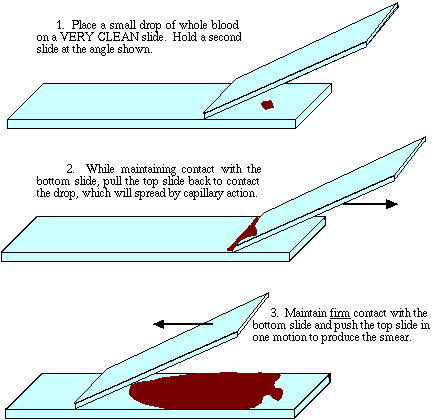
After the smear has dried it can be stained by applying a liberal amount of Wright's stain with a pasteur pipet. Wright's stain contains red and blue dyes that are acidophilic ('acid-loving') and basophilic ('alkaline-loving'), respectively. Avoid skin contact, since most stains are toxic. The stain is allowed to remain on the slide for about 2 minutes, and more is added if it begins to dry up. After a short dip in buffer and/or a rinse in deionized water, the smear is gently blotted dry with bibulous (absorbant) paper. Incidentally, many students have a problem with common sense - my apologies if this insults your intelligence, but DO remove the sheet of bibulous paper from the book before using it for blotting. Don't simply blot the slide onto the entire book.

Microscopic examination of formed elements

No coverslip is needed in order to examine most smears. A proper smear will be quite thick near the location of the original drop and will thin out toward the end. Using 100x bright field magnification, find an area in which the red cells are distributed in one layer, preferably with a slight separation between cells, and pink in color. Darkly stained red cells indicate that the smear was too thick, and cell shape will likely be distorted. They should look like doughnuts, not like stars. You may be able to see the nuclei of white blood cells distributed among the red cells. The nuclei stain a dark blue. Of course, the red cells don't have nuclei.

Because mammalian red blood cells are biconcave they will not appear as uniform disks in the microscope field. Examination at 400x reveals them to be thin in the center. All of the formed elements should be examined at 1000x using the oil immersion technique in order to make positive identifications and to see the fine detail. The drop of oil should be placed directly on the smear.

Most leukocytes (white blood cells) are larger than the erythrocytes, and unless the donor had a nasty blood disease, they will be much more scarce. The two major categories of leukocytes are the granulocytes and the agranulocytes, based on the presence of visible cytoplasmic granules in one type and the absence of visible granules in the other. Each major type is further composed of recognizable sub-types based on staining properties of the granules, size of cell, and proportion of nuclear to cytoplasmic material.

Granulocytes

Granulocytes are also known as polymorphonuclear leukocytes, or "polys" for short. Most have irregular, lobed nuclei. The cytoplasmic granules contain enzymes involved in detoxification of foreign substances, blood clotting, and various immune responses. The neutrophil, the most common granulocyte in humans, is so named because the cytoplasmic granules remain unstained by acidic or basic dyes. Mature neutrophils have a large nucleus with several lobes, although there may be species differences. Neutrophils with one or two lobes are immature or abnormal. The presence of immature leukocytes can indicate a major infection, blood parasites, or a disease such as leukemia. Neutrophils are capable of ameboid motion under some conditions, and as with some other leukocyte types they can accomplish diapedesis, which is the ability to squeeze through the pores of blood vessels to reach a chemotactic source.

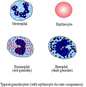
The sex of a blood donor can be determined by examining the neutrophils. Human males have only one copy of the X-chromosome, the genes of which are expressed. In human females, only one X-chromosome in each cell is expressed, while the second is condensed, making the alleles it carries inaccessible. The condensed X-chromosome may jut out as a very small but obvious 'lobe' of the nucleus of the neutrophils, called a Barr body.

Basophils and eosinophils are granulocytes with cytoplasmic granules that stain with basophilic (blue) and acidophilic (red) dyes respectively. Wright-stained basophils are spectacular in appearance, and are usually the rarest of leukocytes. Their granules appear quite large and they stain nearly black, often obscuring the nucleus. The eosinophils, which are often present in large numbers in the presence of a parasitic infection, have numerous red-staining granules.

Agranulocytes

Lymphocytes are smaller than granulocytes, are nearly all nucleus, and typically are nearly as common as neutrophils. The cytoplasm can be seen as a thin area of lighter blue between the nucleus and the cell membrane. Lymphocytes are responsible for the initiation of the immune response and for the acquisition of immunity. Monocytes, the other type of agranulocyte, are much larger than lymphocytes, with a greater ratio of cytoplasm to nucleus. Like neutrophils, monocytes are capable of diapedesis and can respond to chemotactic factors by undergoing hypertrophy (excessive growth) that converts them into macrophages. Macrophages consume foreign substances, such as bacteria, by phagocytosis.

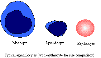
Platelets

The stem cells from which red cells and leukocytes are developed are found in the bone marrow. Also in the bone marrow are giant cells called megakaryocytes. Megakaryocytes disintegrate before enter the bloodstream, so that only the fragments, called platelets, remain. When platelets encounter a damaged surface they swell and stick to the surface and each other to form a plug. The plug seals blood vessels, allowing time for the slower clotting process to take place and form a tight seal. Platelets have no pigmentation, and only lightly stain with Wright's stain, so you may need phase contrast to see them.


Copyright and Intended Use
Visitors: to ensure that your message is not mistaken for SPAM, please include the acronym "Bios211" in the subject line of e-mail communications
Created by David R. Caprette (caprette@rice.edu), Rice University 15 Aug 96
Updated 23 May 05


This page is powered by Blogger. Isn't yours?